The urinary system is responsible for the eliminating of waste and extra fluid in the body, by making and excreting urine.
What is the urinary system and how does it work?
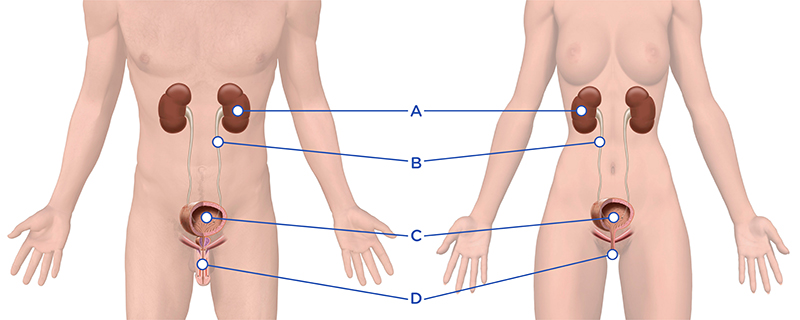
The normal recurring function of emptying and storing of urine is called the micturition cycle. It is divided into a filling and emptying phase. This acts as an interaction between the nervous system and the lower urinary tract. It’s important that all parts of the urinary system work in partnership for normal urination to occur. The urinary system consists of kidneys(A), ureters(B), bladder(C) and urethra(D).
Why is the urinary system important?
Besides filtering and eliminating waste products from the body, the urinary system also maintains our electrolyte balance of sodium, potassium and calcium, blood volume and blood pressure.
What are the different functions of each part of the urinary system?
The kidneys are large, bean-shaped organs located towards the back of the abdomen, either side of the spine. They remove waste products by manufacturing urine. They have a special system of small network of tubes (nephrons) which allow substances to be filtered. The kidneys regulate the amount of water in the body, and humans produce about 1.5 liters of urine a day.
The ureters drain urine from the kidneys to the bladder. They are 25–30 cm long tubes lined with smooth muscle. The muscular tissue uses peristalsis to move urine downwards. The ureters enter into the back of the bladder via the Trigone area, a valve prevents reflux of urine back to the kidneys.
The urinary bladder is a hollow organ which stores urine and located in the pelvis above and behind the pelvic bone. The bladder’s elastic and muscular wall is called the detrusor, and will stretch to hold an average volume of 400-600mls of urine. The inside of the bladder wall is covered with several layers of epithelium (mucosa). The mucosa is folded when the bladder is empty, but stretches out as the bladder fills. It is normal to urinate 4 -8 times per day and (0-1 during the night), normally 200-400 mls each time.
Bladder Trigone
The Trigone is an area inside of the bladder; the triangular area between the ureteric orifices and the internal urethral opening. The area undergoes little change in size during bladder filling. It is very sensitive to stretch as it contains a large number of sensory nerve endings.
Internal Sphincter
A band of the detrusor muscle encircles the urethra at the junction of the bladder to form the internal urethral sphincter.
Urethra and External Sphincter
The urethra is the tube through which urine passes from the bladder to outside the body. The female urethra is around 3-5cm long and almost straight. In males, the urethra is around 20-27 cm and shaped like an S.
The urethra is also an organ of the male reproductive system as it carries sperm out of the body through the penis. An external sphincter is at the furthest end of the urethra and has two important functions; keeping the urine in the bladder, but also relaxes on bladder emptying.
What is the difference between the Internal & External Sphincter?
The external sphincter can, unlike the internal sphincter, be controlled voluntarily. The internal sphincter is under influence of the autonomic (involuntary) nervous system.
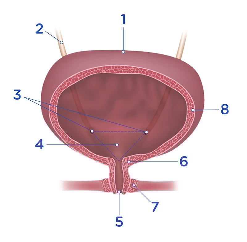
- Urinary bladder
- Ureter
- Ureteral openings
- Trigone
- Urethra
- Internal urethral sphincter (striated muscle)
- External urethral sphincter (striated muscle)
- Detrusor muscle (smooth muscle layers)