
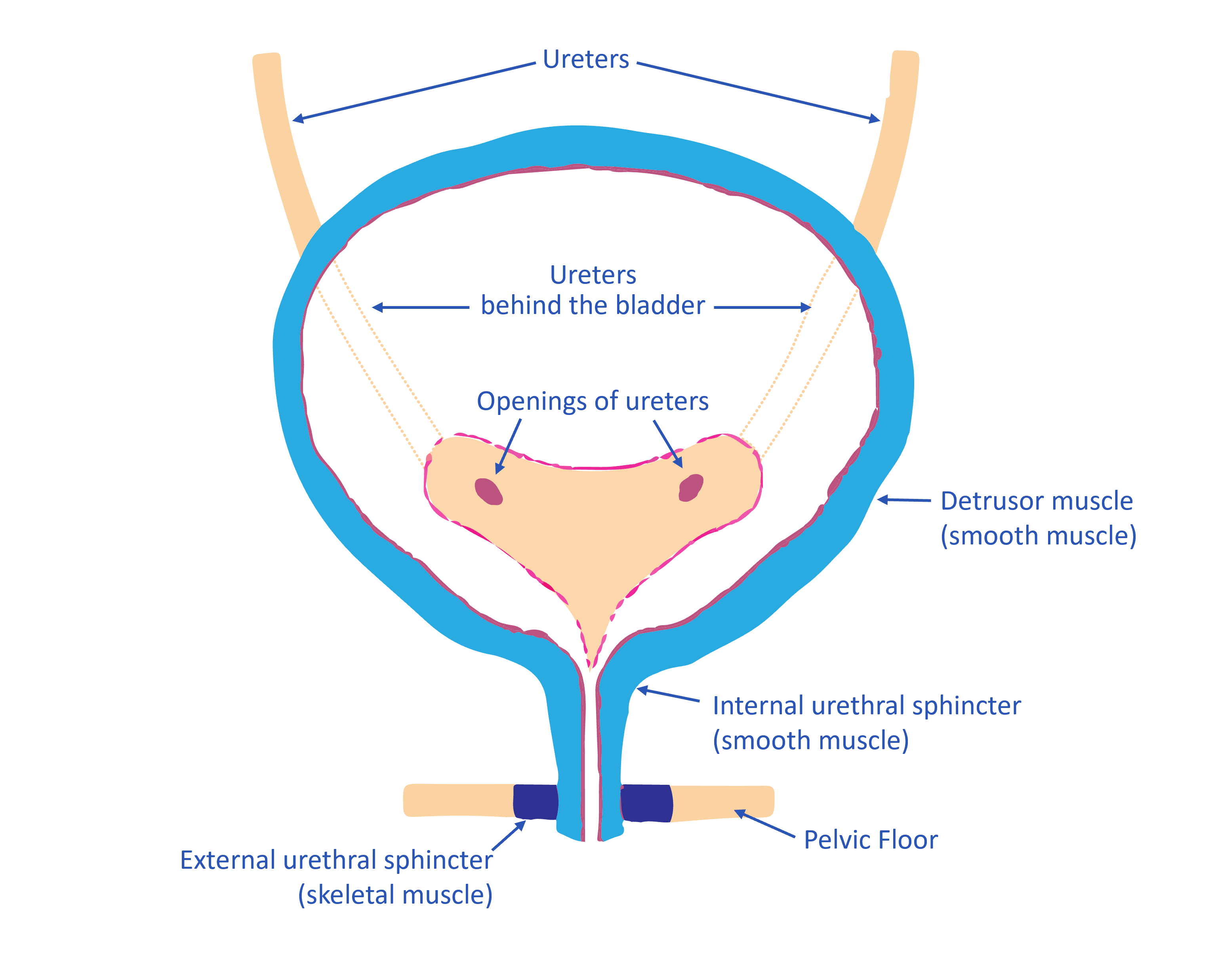
The bladder is a hollow muscular organ in the pelvis, just above and behind the pubic bone. The bladder has two main functions which are the storage and emptying of urine.
It provides continence, by delaying and controlling urination so that the average person urinates four to eight times per day with a bladder volume of 200-400mls (approx.)
Urine is made in the kidneys and travels down two tubes called ureters to the back of the bladder.
The bladder is an elastic organ and is able to increase its volume greatly to accommodate between 600 to 800 ml of urine.
Urination is the process of excreting urine from the bladder.
The central nervous system controls bladder function. Nerve centers for the control of urination are located in the spinal cord, brain stem, and cerebral cortex.
Control of micturition depends on learned behavior during the maturation of the nervous system.
The wall of the bladder consists of the detrusor muscle, which is smooth muscle, and several layers of epithelial mucosa
During urination, the detrusor muscle, contracts, and the urethral sphincters (valves) relax to allow urine to flow out.
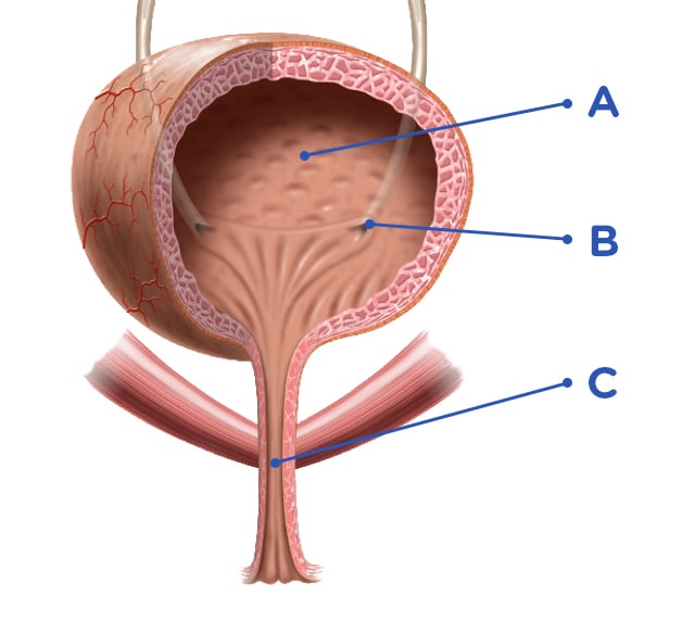
A = Urine Bladder
B = Ureters Opening
C = Urethra
Urine exits the bladder into the urethra, which carries urine out of the body. In females, the urethra is quite short, while in males, the urethra is longer and divided into 3 segments (the prostatic urethra, the membranous urethra, and the penile urethra).
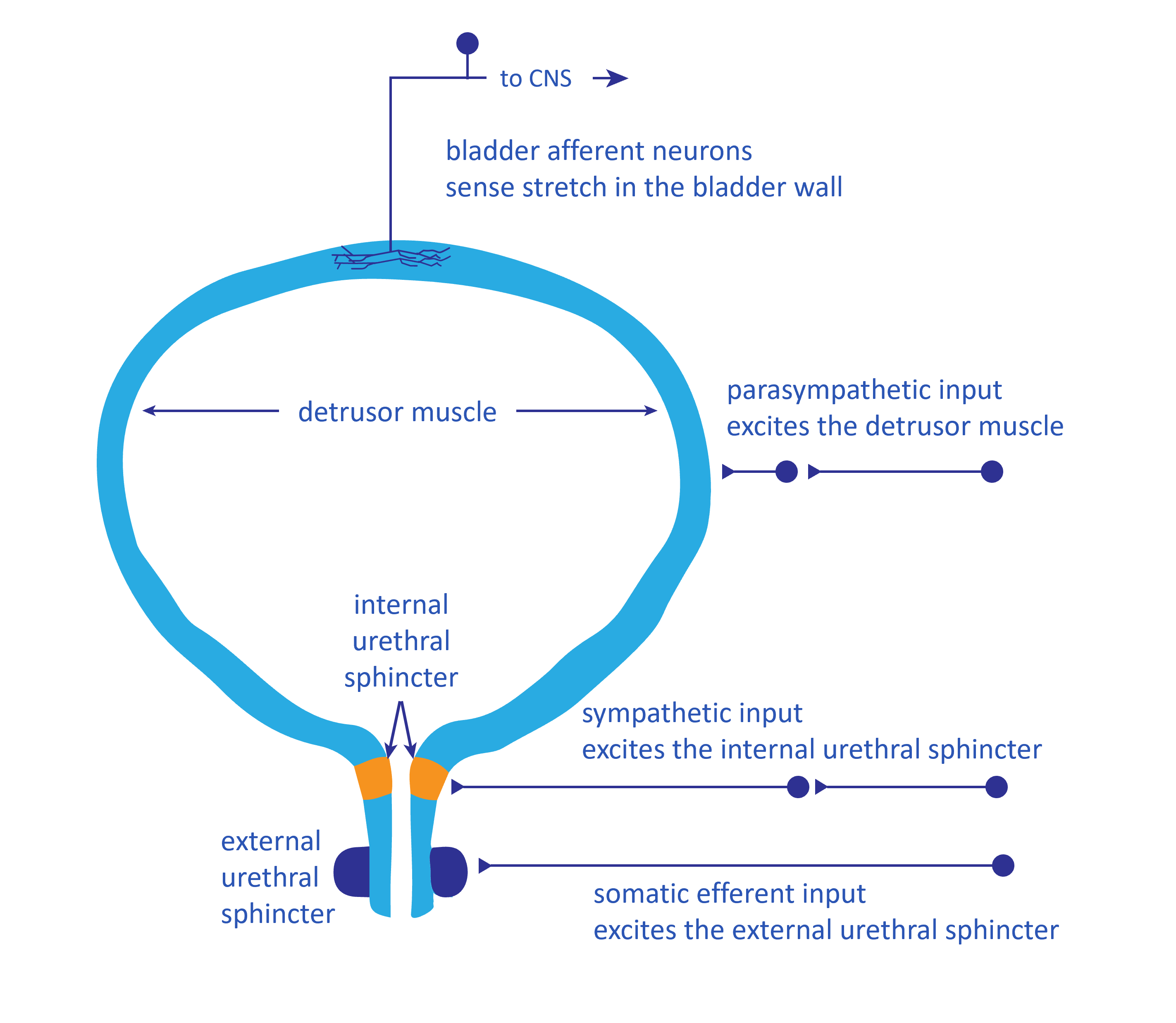
INNERVATION
The bladder is controlled by the central nervous system. The smooth muscles are controlled by the two different divisions of the autonomic nervous system. Parasympathetic inputs stimulate contraction of the detrusor muscle. Sympathetic inputs stimulate contraction of the internal urethral sphincter. Somatic efferent neurons stimulate the contraction of the external urethral sphincter, which is skeletal muscle.
The walls of the bladder stretch as the bladder expands during bladder filling. This is sensed by bladder afferent neurons whose sensory dendrites are located in the bladder wall. Bladder afferents project to the spinal cord and to various regions in the brain that are responsible for coordinating the efferent output to the bladder and urethra.
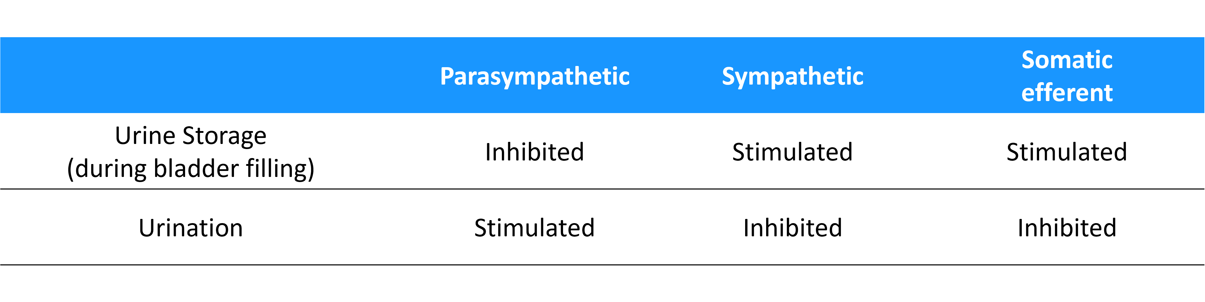
The two different modes of bladder operation
There are two modes of operation for the bladder, which are outlined in the table above: either urine storage or urination. During the time that the bladder is filling, when urine is stored in the bladder, the parasympathetic input to the detrusor muscle is inhibited, and there is the activation of the sympathetic and somatic efferent pathways, causing the two urethral sphincters to contract. The afferent information from the bladder provides a sense of bladder fullness and the urge to urinate. At an appropriate time and place for urination, the parasympathetic efferents become active, causing contraction of the detrusor muscle. At the same time, there is coordinated inhibition of the sympathetic efferents and the somatic efferents so that the internal and external urethral sphincters relax.